How Cutting-Edge Imaging Sheds Light on Microglia and the Fight Against Diabetic Retinopathy
Diabetic retinopathy is a formidable threat, quietly robbing millions of their sight and standing as one of the leading causes of blindness worldwide. Long believed to be caused primarily by damage to the blood vessels in the retina, this form of diabetic eye disease is now being reexamined under the lens of innovative neuroscience. Recent findings from researchers at Kobe University unveil early, hidden signs of the disease, shifting the focus from blood vessels to the retina’s own immune cells and offering hope for new diagnostics and therapies.
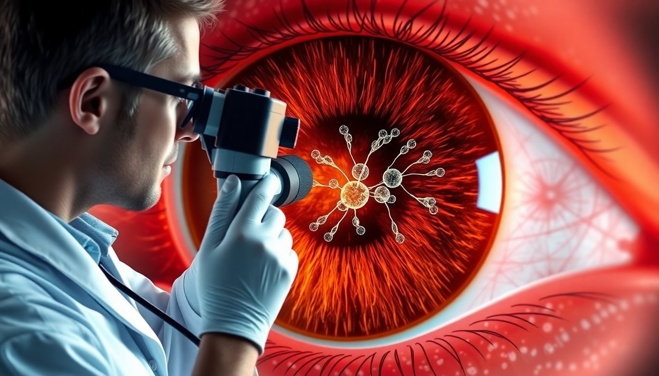
Seeing What Was Once Invisible: The Microglia Connection
For years, scientists have known that high blood sugar resulting from diabetes often leads to damage in the delicate web of blood vessels that feed the retina. This damage was accepted as the principal culprit behind vision loss. But recent studies—especially those spearheaded by Dr. Yoshihisa Tachibana and his team at Kobe University—have revealed a subtler, earlier story: before visible blood vessel damage even occurs, the retina’s own sentinels, called microglia, begin to behave differently.
Microglia act as immune watchdogs in the retina—constantly monitoring their microenvironment, ever vigilant for signs of trouble. These cells are vital first responders, quickly moving to sites of injury or infection and orchestrating inflammation to protect the eye. Yet much of their actual behavior remained a mystery because current technology made it difficult to observe these cells in action within living eyes.
A New Window Into the Living Retina
To crack open this black box, Tachibana’s group engineered an advanced imaging system. Their toolkit included a head-fixation device, custom-made contact lenses, and a specially selected, yet commercially available, objective lens. This creative setup allowed them to peer into the retinas of live diabetic mice with high resolution, tracking the dynamics of microglia over extended periods.
What they found was striking: even before any observable damage to blood vessels, microglia in the retinas of diabetic mice became noticeably more active. Their restless movements indicated they were on high alert, suggesting that inflammation—and not just blood vessel deterioration—might be one of the earliest warning signs of impending vision loss.
“This phenomenon has been overlooked in conventional observation in non-living specimens and is an important finding that provides a new perspective for understanding the pathology of diabetic retinopathy,” notes Tachibana. The realization that microglial hyperactivity may precede visible vascular changes offers not just a deeper understanding, but a potential new arena for medical intervention.
Drug Discovery in Action: Liraglutide’s Surprising Role
Armed with this new imaging technique, the researchers explored more than disease progression—they tested therapies. Diabetic mice were treated with liraglutide, a medication commonly prescribed for diabetes. The results were twofold and unexpected: liraglutide normalized the restless activity of microglia in diabetic mice, but it also subtly calmed microglia in healthy mice. Notably, these effects occurred without any changes to blood glucose levels.
This led to an intriguing discovery—liraglutide appears to act directly on microglia, dialing down their activity through a mechanism unrelated to sugar regulation. For clinicians and researchers, this finding is momentous. It opens the door to possible new treatments for diabetic retinopathy and other retinal diseases, such as glaucoma and age-related macular degeneration, by targeting inflammation at its roots—well before visible damage occurs or vision is irreversibly lost.
A Future of Non-Invasive, Early Diagnosis?
Why does this matter? Early diagnosis is the linchpin of effective treatment for many diseases, including diabetic retinopathy. If doctors could detect abnormal microglial behavior before any visible signs of disease, they could intervene much sooner, potentially preventing blindness altogether. Tachibana envisions just such a future: “We hope that our technology may be put to practical use in clinical settings as a non-invasive diagnostic method, making the eye a window to spotting systemic disease.”
This imaging advancement doesn’t just promise hope for diabetes patients; it lays a foundation for looking at the eye—a uniquely accessible part of the body—as a living atlas for detecting and tracking a range of neurological and systemic diseases. With deeper, real-time insights into cellular behavior, doctors may one day spot not only diabetic retinopathy, but other neurologic and autoimmune challenges, simply by peering into someone’s living retina.
Beyond Diabetes: A Step Forward for Neurotech
The team’s research, conducted in partnership with colleagues from the University of Texas Health Science Center at Houston, the National Institute for Physiological Sciences, and Nagoya University, is also a testament to the power of technological collaboration. By marrying clever engineering with deep biological insight, they’re charting new territory in visual neuroscience—and opening a window, literally and figuratively, into how diseases progress at the earliest, usually hidden, stages.
Given the global burden of diabetes and the looming epidemic of diabetic retinopathy, these findings are a significant stride toward illuminated, effective prevention. More research is needed, of course, to translate this animal model to human eyes and clinical practice. But the promise is palpable: tools and therapies that can intervene before the real damage is done.

Reference
Tachibana, Y., et al. (2024). "Imaging reveals early microglial hyperactivity in diabetic retinopathy and the effects of liraglutide." Proceedings of the National Academy of Sciences (PNAS), Kobe University. https://neurosciencenews.com/diabetes-vision-neurotech-29779/