For years, brain scans for ADHD have produced a confusing mix of results. Now, a clever new method is cutting through the static, revealing consistent structural differences in the brains of children with the disorder.
Attention deficit/hyperactivity disorder (ADHD) affects over five percent of children and adolescents around the globe. It’s a condition characterized by age-inappropriate levels of inattention, hyperactivity, and impulsivity that can create significant challenges in school, social situations, and family life. For decades, neuroscientists have been on a quest to understand the neurological underpinnings of ADHD, using tools like magnetic resonance imaging (MRI) to look for differences in the brains of those with the disorder.
However, this quest has been frustratingly inconclusive. One study might report decreased gray matter volume (GMV) in children with ADHD, while another finds no change, or even an increase. This inconsistency has been a major roadblock, leaving researchers and clinicians with a murky picture. The primary culprit behind these conflicting results isn’t a lack of effort, but a technical one: the MRI machines themselves. Every scanner is slightly different, and comparing data from multiple machines across multiple studies is like trying to compare photographs taken with different cameras under different lighting conditions—the results are noisy and unreliable.
Now, a collaborative team of researchers in Japan has developed and validated a powerful new method to cut through this noise. Their approach, published in Molecular Psychiatry, not only solves the problem of inconsistent data but has also led to one of the clearest discoveries yet about the brain structure in children with ADHD.
The Problem of ‘Noisy’ Data
To conduct large, statistically powerful studies, scientists need to pool data from many different sources. In neuroscience, this means combining MRI scans from various hospitals and research institutions. The problem is that each MRI scanner has its own unique measurement bias. This creates a significant challenge for data harmonization.
Previously, researchers used a statistical method called ComBat harmonization to try and correct for these site-specific differences. While helpful, ComBat has a major drawback: it can be a blunt instrument. In its effort to remove the unwanted ‘noise’ from the machines, it often overcorrects and erases subtle but crucial biological variations among the subjects. It’s like applying a filter to a photo that’s so aggressive it blurs the important details you were trying to see in the first place. This meant that real, underlying brain differences related to ADHD could have been inadvertently wiped out along with the technical errors.
The ‘Traveling-Subject’ Solution
Recognizing this limitation, researchers from the University of Fukui, Chiba University, and The University of Osaka pioneered a more elegant solution: the traveling-subject (TS) method. The concept is both simple and brilliant. Instead of relying on purely statistical corrections, they had a small group of healthy volunteers—the ‘traveling subjects’—get scanned on each of the different MRI machines used in the study.
By scanning the exact same brains on multiple machines, the scientists could create a precise ‘calibration key’ for each scanner. This allowed them to measure the exact bias of each machine and then apply a highly accurate correction to the data from all other participants. In this study, 14 healthy subjects traveled to four different sites over a three-month period, providing the essential data needed to harmonize a much larger, independent dataset.
This TS method proved far superior to the old approach. The results showed that it significantly reduced the measurement bias from the different scanners while, crucially, preserving the natural sampling bias—the real biological variations between individuals that are essential for scientific discovery.

A Clear Signal Emerges: The ADHD Brain Structure
With this powerful new tool in hand, the researchers applied the TS correction to the Child Developmental MRI (CDM) database, which contained scans from 116 children with ADHD and 178 typically developing children. Once the data was ‘cleaned’ of scanner-related noise, a clear and consistent pattern emerged.
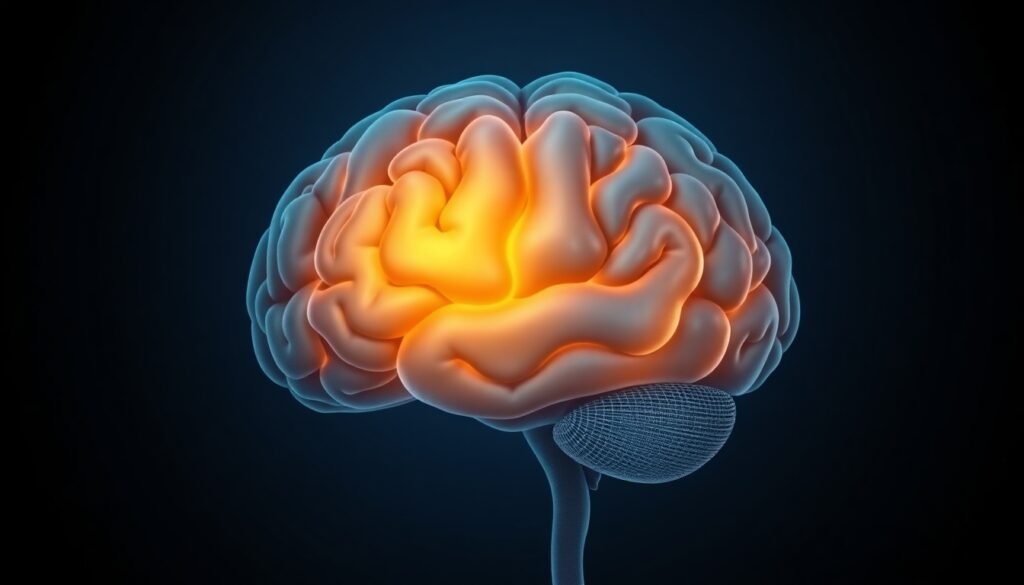
The study revealed that children with ADHD had significantly reduced gray matter volume in the frontotemporal regions of the brain compared to their typically developing peers. These areas, which encompass parts of the frontal and temporal lobes, are hubs for the brain’s executive functions. They are critical for cognitive processes like decision-making, information processing, emotional control, and sustaining attention—the very functions that are most challenging for individuals with ADHD.
Digging deeper, the team identified a particularly significant volume reduction in the right middle temporal gyrus. This specific area is involved in processing external stimuli and social cues. Finding a distinct anatomical difference in a brain region so closely linked to the core symptoms of ADHD provides a powerful physical correlate for the condition.
From Discovery to Diagnosis and Treatment
This breakthrough is more than just an academic achievement; it has profound potential for clinical practice. The ability to reliably harmonize data using the TS method means that future multi-site studies will be more powerful and their findings more trustworthy.
Most importantly, the consistent structural differences found in this study could serve as the first reliable neuroimaging biomarkers for ADHD. A biomarker is a measurable biological indicator of a condition, much like high blood sugar is a biomarker for diabetes. Using brain volume in frontotemporal regions as a biomarker could revolutionize how ADHD is diagnosed and managed.
This could lead to:
- Earlier, More Objective Diagnosis: Current ADHD diagnosis relies heavily on behavioral observations and checklists. An objective biomarker could supplement these methods, allowing for earlier and more accurate identification of the disorder.
- Personalized Interventions: By understanding the specific neuroanatomy of a child’s brain, clinicians could one day tailor treatments—whether behavioral therapy, cognitive training, or medication—to their unique biological profile.
- Effective Treatment Monitoring: Follow-up scans could provide objective data on whether an intervention is having the desired effect on brain development and structure, allowing for more responsive and effective care.
As the study’s authors conclude, this approach aims to facilitate earlier diagnosis and more precise, individualized interventions. In the long term, this could dramatically improve the quality of life for children with ADHD and reduce their risk of developing secondary psychiatric disorders like anxiety and depression.
This research represents a significant leap forward, cutting through years of confusing data to provide a clearer picture of the ADHD brain. It paves the way for a future where this common neurodevelopmental disorder is understood, diagnosed, and treated with a new level of scientific precision.
Reference
Shou, Q., Mizuno, Y., Hirano, Y., & Kagitani-Shimono, K. (2025). Brain structure characteristics in children with attention-deficit/hyperactivity disorder elucidated using traveling-subject harmonization. Molecular Psychiatry.