Subtitle: Exploring How the Brains of People Without Visual Imagery Respond Differently During Mental Tasks
The ability to conjure images in the mind—whether retracing a childhood memory or envisioning a scene described in a book—is often taken for granted. Yet, for individuals with a condition known as aphantasia, visual mental imagery is a blank slate: they cannot voluntarily generate images in their mind’s eye. As neuroscience grapples with the puzzle of aphantasia, a new study sheds light on the distinctive ways these brains process information, thanks to recordings of electrical activity during mental tasks.
What Is Aphantasia?
Aphantasia is not a lack of imagination, but rather a lack of voluntary visual mental imagery. First described in depth in the mid-2010s, scientists and the public alike have shown keen interest in understanding its broader implications—not only for memory and creativity, but also for how the brain represents and manipulates information.
The condition appears to be present in about 1-3% of the population, yet until recently, much about its neural mechanisms remained unknown. Is the absence of visual imagery just a quirk of subjective experience, or is it reflected in genuine differences in how the brain processes information?
The Role of EEG in Unlocking Brain Mysteries
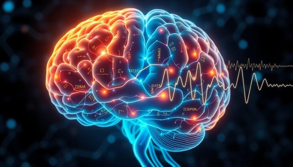
Electroencephalography, or EEG, offers a non-invasive window into the brain’s electrical activity. By placing sensors on the scalp, researchers can record the fleeting waves and patterns generated as different areas process information. Of particular relevance in cognitive neuroscience are event-related potentials (ERPs): brief changes in the EEG signal that occur in response to specific mental tasks or sensory stimuli.
Certain components of the EEG, such as the P300 wave or oscillations in the alpha and theta bands, are known correlates of attention, memory, and imagery. By comparing these signals across individuals with and without aphantasia, researchers hope to identify the neural signatures unique to each group.
A New Study: Tasks, Trials, and Revealing Contrasts
A recent study led by Katherine Boere and colleagues took on this challenge. The research team recruited both individuals with aphantasia and those with typical imagery abilities. Participants completed a battery of working memory and imagery tasks—all while their brain activity was meticulously tracked using EEG.
Crucially, the tasks in this experiment were designed to tap into the sorts of mental activities where imagery might play a role, such as recalling or manipulating visual information. Examples include remembering the layout of objects or mentally rotating shapes—classic challenges in cognitive psychology.
What Did the EEG Reveal?
The EEG data revealed striking differences in how aphantasic and non-aphantasic brains processed the same tasks. For example, the amplitude and timing of the P300 wave—a marker often linked with decision-making and stimulus evaluation—differed between groups. These distinctions weren’t just statistical quirks; they reflect genuine divergences in cognitive strategy and neural recruitment.
Individuals with aphantasia showed altered activity in brainwave oscillations, particularly in the delta and theta frequency bands. The delta band, associated with internal attention and cognitive processing, seemed to be engaged differently during mental tasks in the aphantasic group. Theta oscillations, known to increase with memory load, also showed unique patterns.
Importantly, the study’s findings support the idea that while aphantasics can perform visual working memory tasks just as accurately as others, they may do so using different strategies—relying less on mental images and perhaps more on verbal or analytical representations.
Rethinking Mental Imagery and Cognitive Diversity
This growing body of evidence helps refine our understanding of how the brain handles visual information in diverse ways. Rather than seeing aphantasia as a simple deficit, it may be better viewed as a variant in cognitive processing—an alternative route through the mental landscape.
Previous studies have shown that people with aphantasia:
- Can perform mental rotation tasks, but often do so more slowly or with different strategies
- Might show reduced vividness in recalling past events
- Display characteristic differences in pattern connectivity between brain regions such as the hippocampus and occipital cortex
The EEG study adds a dynamic, real-time element to this story: the brain is flexible, and when imagery isn’t available, it finds ways to compensate—sometimes visible in the very waves of neural activity.
Questions and Considerations for Future Research
While this study significantly enhances our grasp of aphantasia’s neurobiology, much is left to explore. Do these EEG differences appear in other types of mental tasks? How do they relate to emotional processing or learning? Could there be subtypes of aphantasia, with differing compensatory mechanisms?
One exciting aspect of EEG research is its accessibility: portable, relatively low-cost devices bring the prospect of wider-scale studies, including in populations outside of traditional academic settings. This could accelerate discoveries around aphantasia and other conditions with subjective and elusive symptoms.
The Value of Cognitive Diversity
In a world that celebrates creativity and out-of-the-box thinking, the study of aphantasia serves as a reminder that there are many paths to a similar goal. The brain’s adaptability is on full display, challenging us to rethink what is ‘normal’ in mental experience.
Ultimately, understanding the neural basis of aphantasia not only deepens our knowledge of mental imagery but also broadens the conversation about cognitive diversity and the ever-mysterious landscape of the human mind.
Reference
Boere, K., Krempel, R., Walsh, E. et al. (2025). Task evoked EEG reveals neural processing differences in aphantasia. Scientific Reports. https://doi.org/10.1038/s41598-025-27735-x