Understanding network-specific changes in the brain’s main bridge and their implications for aging minds
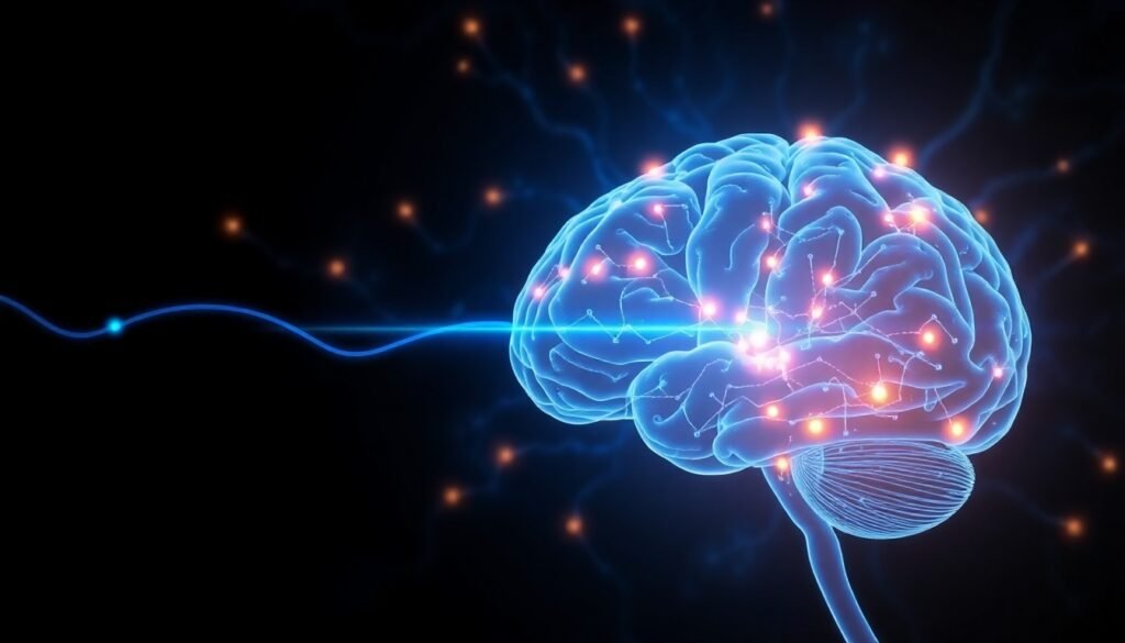
The human brain is famously complex, with two hemispheres working together through a bundle of nerve fibers called the corpus callosum. As we age, this central connector changes—but not always in the same way across different brain networks. Recent research sheds new light on how aging affects specific pathways within the corpus callosum and how these shifts may influence cognition later in life.
The Unsung Hero: Corpus Callosum’s Role in Brain Communication
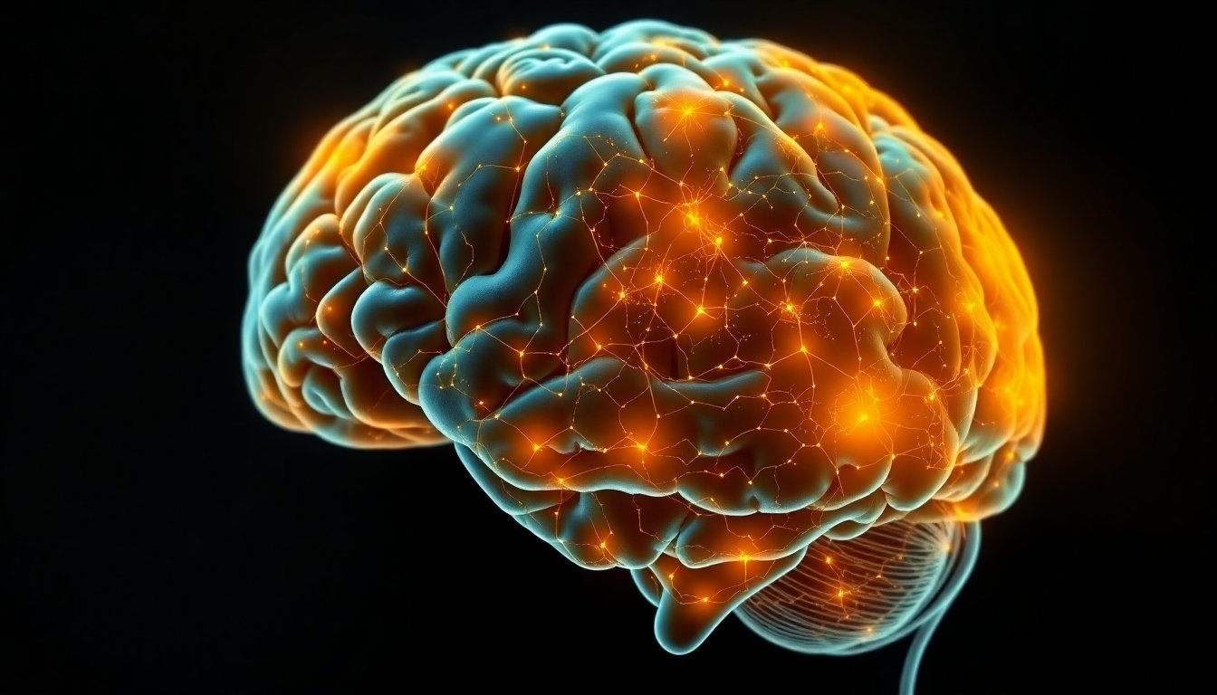
The corpus callosum—an arching band of white matter—serves as the largest highway for interhemispheric communication. It enables the left and right sides of our brains to share and integrate information, allowing for coordinated perception, movement, and thought. Decades of neuroscience research, from early anatomic studies to advanced brain imaging, have emphasized its role in balancing excitation and inhibition between hemispheres and supporting cognitive functions like attention, memory, and problem-solving.
A New Lens on Brain Aging
While it’s known that the corpus callosum changes across the lifespan, the latest study led by Mohammad Hadi Aarabi (2025) at the University of Padova dives even deeper. Their approach—called tract-to-region analysis—allowed them to examine how different connections within the corpus callosum, specific to distinct brain networks, evolve with age.
Traditionally, research focused on global measures of the corpus callosum’s size or thickness. However, these broad-brush approaches miss the nuances of network-level connectivity—the idea that particular regions of the corpus callosum link specific functional areas of the brain, each supporting distinct types of cognition.
What Did the Study Find?
Network-Specific Aging Patterns
Not all parts of the corpus callosum age equally. This study revealed that:
- Certain tracts—bundles of nerve fibers linking specific cortical networks—showed greater age-related declines in integrity than others.
- These declines weren’t uniform; the way the corpus callosum changes depends on which areas and networks it connects. For example, connections supporting executive functions (like planning and flexible thinking) may deteriorate differently from those involved in sensory processing.
Cognitive Implications
Perhaps most intriguing, these structural changes weren’t just anatomical curiosities—they had observable consequences for cognition:
- Older adults showed network-specific associations between callosal integrity and cognitive abilities. That is, declines in certain tracts were more strongly linked to reduced cognitive performance than others.
- The impact of aging on cognition appears, in part, to depend on which brain networks are most affected by callosal changes.
This nuanced understanding may explain why some older adults experience declines in certain cognitive domains while others remain relatively sharp; the underlying network-specific brain pathways may be aging at different rates for different individuals.
The Bigger Picture: Evolution and Development
The corpus callosum is not unique to humans but is especially well-developed in our species compared to other primates. Its evolution and development have played a crucial role in the emergence of complex cognition, language, and social behaviors. Throughout adulthood and aging, its integrity supports efficient communication and functional lateralization—the specialization of each hemisphere for certain tasks.
Comparative studies in chimpanzees and baboons underscore that while age-related changes occur across species, the complexity and variability in human corpus callosum networks may contribute to our unique cognitive trajectories with aging.
Technology Meets Neuroscience
The tract-to-region method harnessed advanced brain imaging and connectome mapping—techniques that allow researchers to chart the brain’s wiring at an unprecedented scale. By pairing these methods with large-scale lifespan datasets, like those compiled by the Human Connectome Project, scientists can clarify how diverse brain networks are impacted by aging and which changes have meaningful effects on daily functioning.
Why Does This Matter?
Understanding which parts of the brain’s main bridge age earliest and how this impacts cognition is more than academic. It can help:
- Target interventions to slow or compensate for specific neural changes as we age.
- Refine our grasp of normal versus pathological aging and distinguish between healthy decline and disease.
- Guide future research into aging-related disorders affecting connectivity—such as stroke, dementia, and multiple sclerosis.
The Road Ahead: Personalized Brain Health
As new connectivity mapping tools emerge, we move closer to precision neuroscience:
- Identifying individualized patterns of brain aging may one day allow doctors to tailor cognitive training or interventions based on a person’s unique neural profile.
- By focusing on network-specific changes, therapies could be directed not just at the brain globally, but at the most vulnerable pathways within each person.
Final Thoughts
The architecture of our brains—and how it changes with age—underpins everything from memory to creativity. By peering into the intricate connections of the corpus callosum, researchers are illuminating the pathways that make us who we are, and helping us chart a course for healthy brain aging.
References:
Aarabi, M.H. (2025). Network-specific corpus callosum aging and age-moderated cognitive associations using tract-to-region analysis. Communications Biology. https://doi.org/10.1038/s42003-025-09219-w
Additional sources cited within the article from: Bloom & Hynd (2005); Syrmos et al. (2023); Thiebaut De Schotten & Forkel (2022); Danielsen et al. (2020); Delvenne & Malloy (2025); Westerhausen et al. (2021); Martins-Costa & Knoblich (2023); and others as acknowledged in the study.