Subtitle: Scientists engineer functional brain-like tissue without animal products, opening new doors to neuroscience research and drug discovery.
Introduction
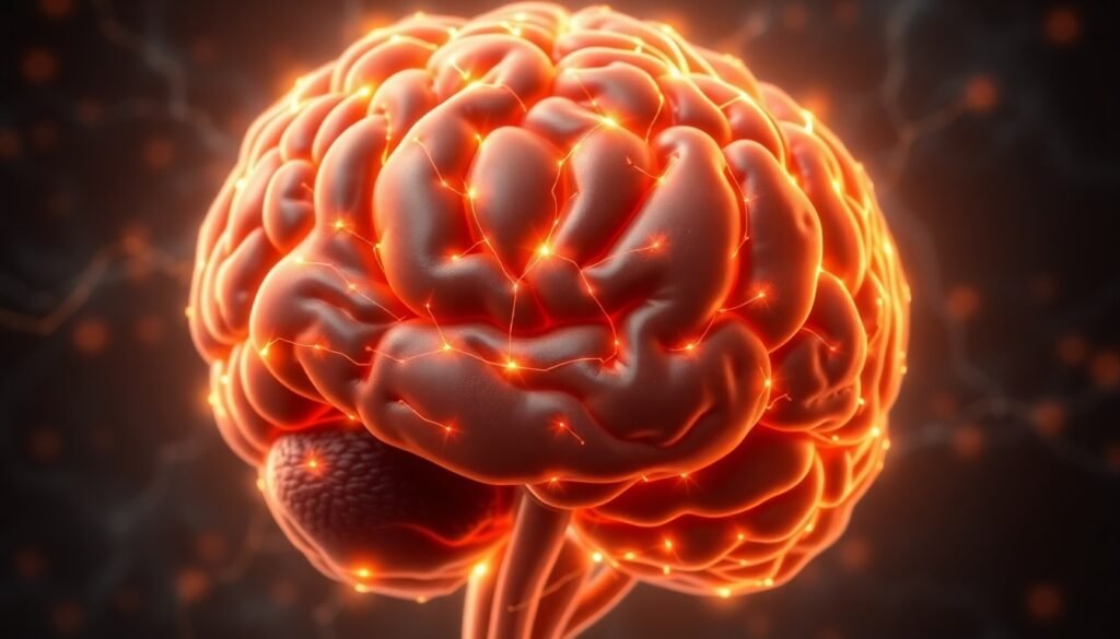
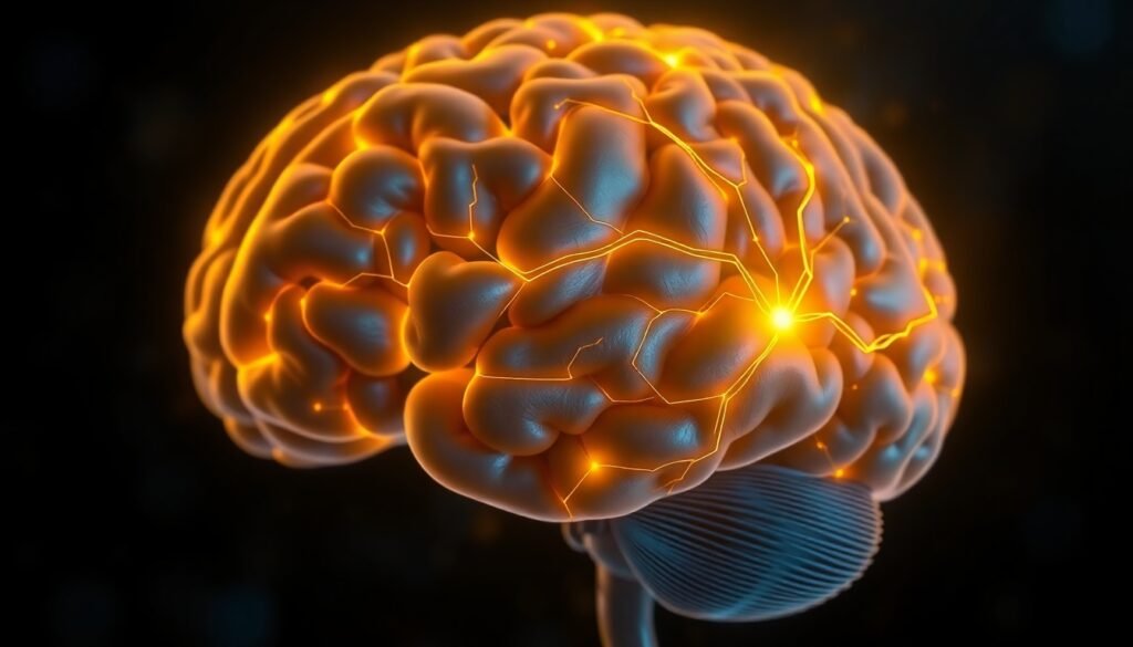
The intricate mysteries of the human brain have always intrigued scientists, yet the ethical and practical hurdles of studying brain tissue in the lab have limited progress. Traditionally, researchers have relied heavily on animal-derived materials or even actual animal brains to grow and test brain-like tissue. Not only does this raise ethical questions, but models built on these animal components often lead to results that don’t translate well to humans because of significant genetic and physiological differences. Now, a breakthrough from researchers at the University of California, Riverside, promises to revolutionize the field: for the first time, scientists have engineered functional, brain-like tissue using a completely animal-free method.
The Breakthrough: Building a Brain Without Animals
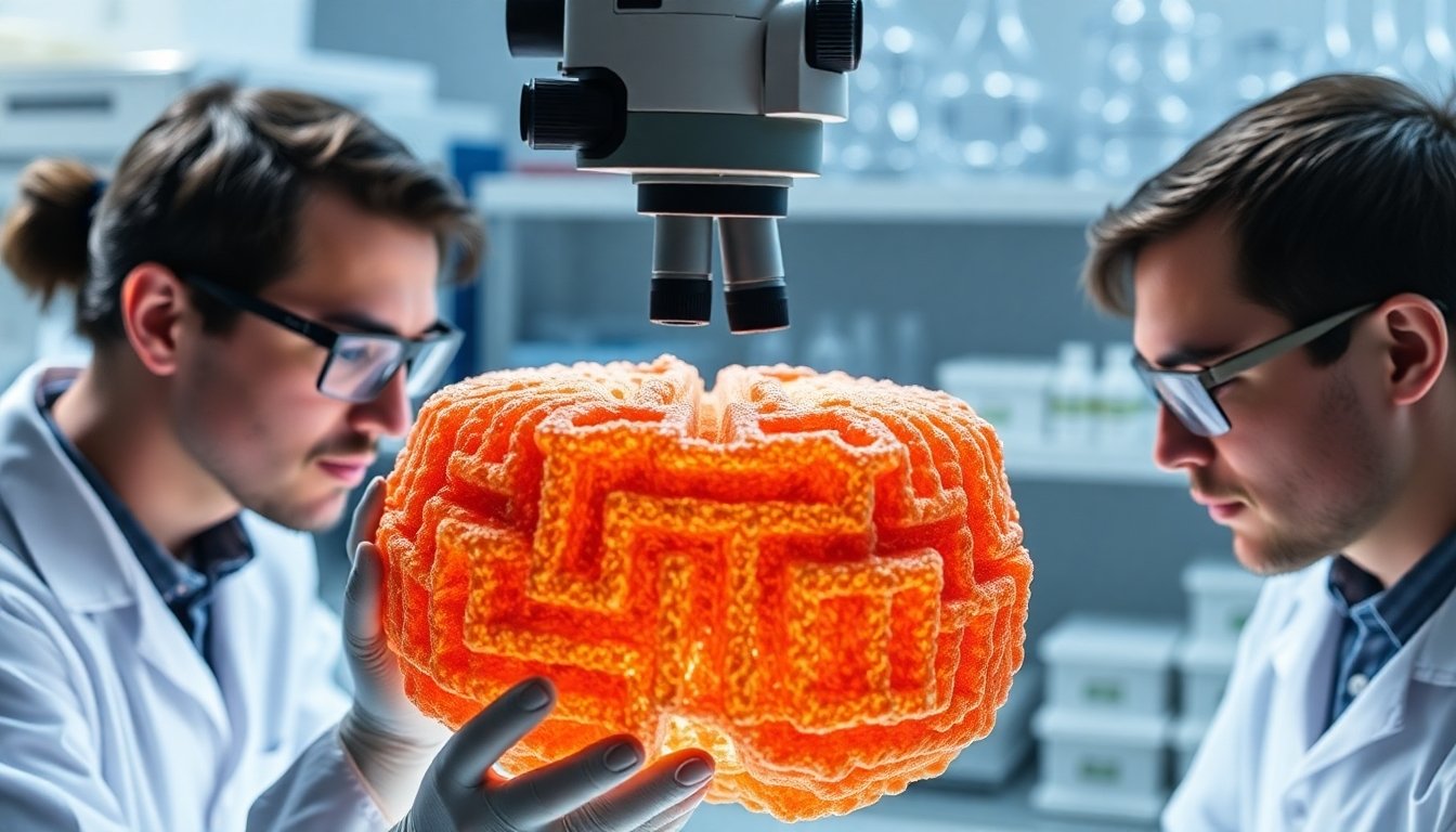
At the core of this innovation is a common polymer—polyethylene glycol (PEG)—previously known for its chemical neutrality and, until now, its inability to support living cells on its own. The research team, led by Iman Noshadi, an associate professor of bioengineering at UC Riverside, transformed PEG into a scaffold of porous, maze-like channels. This structure is reminiscent of the complex environments in which brain cells thrive and communicate.
Importantly, this new scaffold eliminates the need for animal-derived proteins such as laminin or fibrin, which have traditionally been used as coatings to help cells stick, grow, and organize. While these proteins made cell cultures possible in the past, their composition is often variable and poorly defined, making it difficult to reproduce experiments reliably. By removing these materials from the equation, the PEG-based platform offers unprecedented consistency and ethical integrity.
How Does It Work?
The technology hinges on a clever fabrication process: the team used a mixture of water, ethanol, and PEG, channeled through concentric glass capillaries. As this mixture hit an outer stream of water, its components began to separate. A sudden flash of light stabilized the resulting separation, locking the structure into a highly porous, interconnected network.
This unique architecture allows oxygen and nutrients to flow freely throughout the tissue, ensuring that the donated stem cells placed into the scaffold are well-nourished and capable of growing into healthy, mature neural clusters. As they develop, these cells form networks that more closely mimic the organization and function of real human brain tissue.
A Step Toward Better Drug Testing and Disease Modeling
One of the biggest motivations behind this animal-free brain tissue is the urgent need for better models in drug testing and disease research. The U.S. Food and Drug Administration (FDA) has even begun encouraging a move away from animal testing requirements in drug development, recognizing both the ethical challenges and the limitations of animal-based models.
With this PEG-based scaffold, researchers can grow patient-specific neural tissues that display genuinely human activity. This opens the possibility of testing experimental drugs in cultures that reflect a patient’s unique neural makeup, taking personalized medicine to new heights. Furthermore, because the platform is stable and chemically defined, these cultures can survive and mature over longer time frames—a major advantage for studying slow-developing neurological conditions like Alzheimer’s disease or the effects of traumatic brain injuries.
Prince David Okoro, the study’s lead author and a doctoral candidate in Noshadi’s lab, highlights the significance of this feature: "Since the engineered scaffold is stable, it permits longer-term studies. That’s especially important as mature brain cells are more reflective of real tissue function when investigating relevant diseases or traumas."
Towards a Multi-Organ Future
For now, the scaffold is about two millimeters wide, but the team is actively working to scale up the technology. They are also expanding their approach to other tissues, having already submitted related work on liver models. The ultimate vision is an interconnected suite of organ-level systems—a kind of miniaturized, lab-grown body—that enables scientists to see, in real-time, how various organs react to drugs or disease and how problems in one tissue might affect another.
Such an integrated platform would represent a massive leap in both the complexity and ethical standing of biomedical research. It could also significantly accelerate the path from lab discoveries to real-world therapies, reducing or even eliminating the need for animal testing altogether.
Implications and Future Prospects
This animal-free, PEG-based neural scaffold is more than a technical achievement. It’s a statement about the direction of science: towards greater rigor, reproducibility, and ethical responsibility. By offering a reliable, scalable, and humane platform for growing brain tissue, this research lays the groundwork for more accurate drug testing, richer disease models, and ultimately, better treatments for neurological disorders.
As the technology matures and scales, it may soon become a staple in neuroscience labs around the world—heralding a future where medical research is both cutting-edge and conscientious.
Reference
Noshadi, I., Okoro, P. D., et al. (2024). Bicontinuous microarchitected scaffolds provide topographic cues that govern neuronal behavior and maturation. Advanced Functional Materials.