A groundbreaking imaging technique reveals that the impact of a spinal cord injury extends far beyond the spine, offering new hope for diagnosis and treatment.
Every year, tens of thousands of people face the life-altering diagnosis of a traumatic spinal cord injury (SCI). In the United States alone, it’s estimated that over 300,000 people live with the consequences, which can range from chronic pain to a partial or complete loss of sensation and movement. For doctors and patients alike, one of the greatest challenges has been understanding the full extent of the damage. Traditional imaging methods like X-rays and CT scans are excellent at showing structural damage—a fractured vertebra, for instance—but they can’t see the invisible wound: the disruption to the intricate network of nerves and synapses that carry life’s essential signals.
This diagnostic gap has been a major roadblock. Without a clear picture of the underlying neural damage, it’s difficult to predict a patient’s recovery, monitor their progress, or determine if a new therapy is truly working. But now, a team of researchers from the Yale School of Medicine has developed a revolutionary approach that pulls back the curtain on this hidden damage. Using an innovative PET tracer, they have found a way to map synapse loss not only at the site of the injury but also in distant regions of the brain, revealing for the first time the widespread neurological fallout of an SCI.
Lighting Up the Synapses
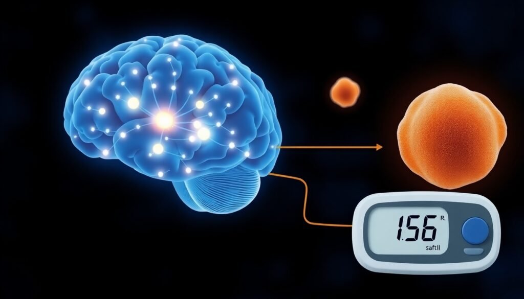
The breakthrough hinges on a powerful imaging technique called Positron Emission Tomography (PET) and a newly developed radiotracer known as [18F]SynVesT-1. In simple terms, a PET scan works by detecting a special radioactive molecule—the tracer—that is designed to travel through the body and bind to a specific target. In this case, the target is a protein called synaptic vesicle glycoprotein 2A, or SV2A.
SV2A is a crucial component of synapses, the vital connections between neurons that allow them to communicate. Think of synapses as the microscopic wiring that makes up the entire central nervous system. The [18F]SynVesT-1 tracer is essentially a synapse-seeking missile. When injected, it travels to the brain and spinal cord and latches onto SV2A. The PET scanner can then detect the tracer’s signal, creating a detailed map of synaptic density. The brighter the signal, the more healthy synapses are present; a weaker signal indicates synapse loss.
To test their new tool, the researchers used a rat model of spinal cord injury. They induced a moderate contusion injury in the mid-thoracic region of the spine (T7) in one group of rats and performed a sham procedure on a control group. They then performed PET scans on day one after the injury and again between days nine and eleven.

The Ripple Effect: An Injury’s True Reach
The results were both confirming and astonishing. As expected, the PET scans revealed a dramatic loss of synapses directly at the injury’s epicenter. Compared to the healthy control rats, the injured rats showed a 58% reduction in the tracer’s signal on day one, a figure that remained high at 52% by day eleven. This finding alone was a powerful proof-of-concept, demonstrating that [18F]SynVesT-1 PET could effectively and quantitatively measure the immediate neural damage from an SCI.
But the most profound discovery came when the researchers looked beyond the spine. The scans revealed that the injury’s impact was not a localized event. The rats with spinal cord injuries also showed significant synapse loss in distant and seemingly unconnected areas of the brain, including the amygdala and the cerebellum. The amygdala is a key hub for processing emotions, while the cerebellum is critical for coordinating movement and balance. This finding highlights that an injury to the spinal cord sends shockwaves throughout the entire central nervous system.
To validate these surprising results, the team used other advanced techniques. Ex vivo diffusion tensor imaging (DTI), a type of MRI that maps the brain’s white matter tracts, confirmed that there was corresponding fiber damage in other brain regions, such as the internal capsule and somatosensory cortex. This multi-pronged approach provided compelling evidence that an SCI triggers a cascade of neurodegeneration that reaches far into the brain.
From the Lab to the Clinic: A New Era for SCI Care
While this research was conducted in animal models, its potential clinical implications are immense. As associate professor and lead researcher Dr. Jason Cai explains, “There is an urgent need for a quantitative and non-invasive imaging method for neural network changes after spinal cord injury.” This new SV2A PET imaging technique appears to be exactly that.
For the first time, doctors may have an objective tool to see what truly matters: the health of the neural connections. This could transform how spinal cord injuries are managed in several key ways:
- More Precise Diagnosis and Prognosis: By quantifying the extent of synapse loss in both the spine and brain, clinicians could gain a much clearer understanding of an individual’s injury, potentially leading to more accurate predictions about their long-term recovery.
- Objective Monitoring: Doctors could use serial PET scans to track changes in synaptic density over time, providing a clear, biological metric of whether a patient is recovering or degenerating.
- Accelerating New Therapies: This technique offers a powerful way to test the effectiveness of novel treatments. Researchers could objectively measure whether a new drug or rehabilitation strategy is successful at protecting or even regenerating synapses, dramatically speeding up the development of effective therapies.
Ultimately, this could pave the way for more personalized medicine. By understanding the unique pattern of synaptic damage in each patient, doctors could tailor therapeutic strategies to their specific needs. “Our work has potential to revolutionize the way spinal cord injury is diagnosed and monitored in the clinic,” Cai noted.
This study marks a significant leap forward in our ability to visualize the complex consequences of spinal cord injury. By revealing the hidden, system-wide impact on the nervous system, this innovative PET imaging technique provides a critical new tool for researchers and a new beacon of hope for patients and families navigating the difficult journey of recovery.
Reference
Cai, J., et al. (2024). [18F]SynVesT-1 PET Detects SV2A Changes in the Spinal Cord and Brain of Rats with Spinal Cord Injury. The Journal of Nuclear Medicine.