A groundbreaking study finds that the shape of brain grooves in former football players differs from non-athletes, offering a potential new path for diagnosing the devastating neurodegenerative disease before it’s too late.
Chronic Traumatic Encephalopathy, or CTE, casts a long shadow over contact sports. It’s a progressive and devastating neurodegenerative disease linked to repeated head impacts, yet it holds a frustrating secret: it can only be definitively diagnosed after death. For living athletes, families, and clinicians, this has meant navigating a world of uncertainty, trying to manage symptoms without a clear diagnosis. But a new study, led by an international team of researchers including those at NYU Langone Health, offers a significant glimmer of hope, suggesting we may be able to see the earliest footprints of CTE in the living brain.
By analyzing MRI scans, the researchers have identified subtle, yet significant, structural differences in the brains of former football players compared to their peers who never played contact sports. These differences are not random; they appear in the very regions known to be affected in postmortem-confirmed cases of CTE, providing a potential roadmap for early detection.

The Landscape of the Brain
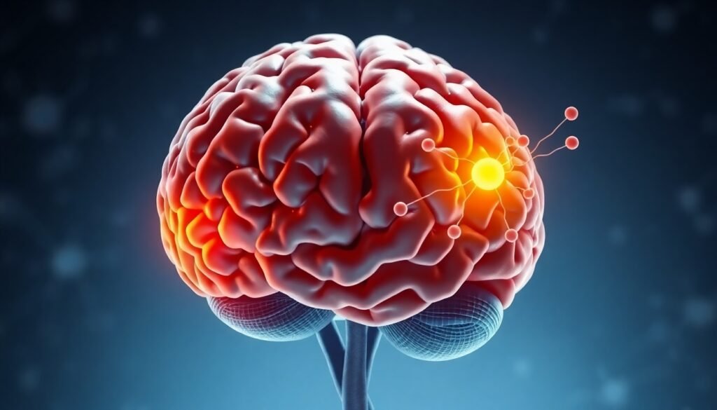
To understand the study’s findings, we need to look at the brain’s intricate topography. The outer layer of the brain, the cerebral cortex, is folded into a complex pattern of ridges (gyri) and grooves (sulci). These folds are not just for show; they dramatically increase the brain’s surface area, packing more cognitive power into the limited space of the skull. In CTE, autopsies have revealed a telltale sign: a buildup of an abnormal protein called tau, particularly in the depths of these sulci, often near blood vessels. This accumulation leads to the death of brain cells and, over time, the cognitive, behavioral, and mood changes associated with the disease.
The research team hypothesized that if tau pathology starts deep within these grooves, it might eventually cause cortical atrophy—a shrinking of the brain tissue—that would alter the shape of the sulci themselves. Could these changes, perhaps a shallowing or widening of the grooves, be visible on a high-resolution MRI scan?
To find out, they analyzed brain scans from 169 former college and professional football players and compared them to scans from 54 carefully matched men of similar age, weight, and education who had no history of playing contact sports or serving in the military. The football group included players in high-impact positions like linemen, receivers, and defensive backs, who had played for at least six years at the college level or twelve years professionally. Quarterbacks were excluded due to their relatively lower exposure to head trauma.
Telltale Differences in the Grooves
The results were striking. The study, published in the journal Brain Communications, revealed two key findings. First, the former football players, on average, had shallower left superior frontal sulci. This is a major groove located in the front, left side of the brain—a region that past autopsy studies have consistently shown is one of the first places affected by CTE pathology. This finding provides a direct, structural link between the changes seen in living players and the known pathology of the disease.
Second, the researchers found a compelling “dose-response” relationship. The more years a person had played football, the wider their left occipitotemporal sulci—a groove on the left side of the brain—tended to be. This correlation strengthens the argument that the physical demands and repeated head impacts of the sport are directly contributing to these measurable changes in brain structure.
“Our study shows what we believe can be the first structural differences that tell apart brains more at risk of developing chronic traumatic encephalopathy from the brains of people who are less at risk,” explained study senior investigator Hector Arciniega, PhD, an assistant professor at the NYU Grossman School of Medicine. “The work also proves that we can apply what we know about the physical changes observed postmortem in the brains of those with confirmed chronic traumatic encephalopathy to brain scans of living people at increased risk for it.”
The Path Toward a Living Diagnosis
These findings represent a critical step toward developing an in vivo biomarker for CTE—an objective, measurable indicator of disease risk in a living person. Identifying individuals at high risk before symptoms become severe could revolutionize how the disease is managed. It opens the door for future therapies to be administered at a stage when they might be able to slow or even halt the disease’s progression, before the damage becomes irreversible.
However, the researchers are careful to temper excitement with scientific caution. This is not a diagnostic test for CTE—at least, not yet. For one, it remains unclear why the differences were detected only on the left side of the brain. Furthermore, the observed sulcal changes did not correlate with the participants’ performance on psychological tests for memory and learning, nor did they align with levels of tau protein detected by current PET imaging techniques. This highlights the immense complexity of CTE and suggests that sulcal morphology is likely one piece of a much larger puzzle.
Dr. Arciniega and his team emphasize that a reliable clinical test is still years away. The next steps will involve validating these findings in larger, more diverse groups and expanding the investigation to include athletes from other contact and collision sports like soccer, hockey, and boxing. The ultimate goal is to combine multiple biomarkers—including imaging, fluid analysis, and clinical assessments—into a comprehensive CTE risk profile.
While the finish line is not yet in sight, this research marks a significant advance. For decades, CTE has been a ghost in the machine, its presence only confirmed after it has already run its course. By learning to read the subtle language of the brain’s own geography, scientists are moving closer than ever to shining a light on the disease, offering a future where diagnosis, prevention, and treatment are a reality for living athletes.
Reference
Arciniega, H., Jung, L., Mirmajlesi, A., Stearns, J., Heller, C., Im, B., … & Koerte, I. K. (2023). Sulcal morphology in former American football players. Brain Communications, 5(5), fcad261. https://doi.org/10.1093/braincomms/fcad261