New research reveals a specific group of brain cells that work while you sleep, turning fat into fuel to keep your blood sugar stable—a discovery that could unlock new insights into prediabetes.
As you settle into sleep each night, your body enters a state of fasting. For the next several hours, without any incoming food, it must perform a delicate balancing act: ensuring that your brain and other vital organs receive a steady supply of energy. The primary fuel for this is glucose, or blood sugar. A severe drop in glucose, a condition known as hypoglycemia, can be dangerous. So, how does your body prevent this from happening night after night? The answer, it turns out, lies deep within the command center of your body: the brain.
For decades, scientists have understood that the brain plays a crucial role in managing blood glucose during stressful situations, like prolonged fasting or emergencies. However, its role in the routine, day-to-day maintenance of our metabolic health has been less clear. A groundbreaking study from the University of Michigan, published in the journal Molecular Metabolism, has illuminated a previously hidden network of brain cells that acts as a silent guardian, working diligently through the night to maintain this crucial balance.
The Brain’s Metabolic Thermostat
At the heart of this discovery is a small but powerful region of the brain called the hypothalamus. Nestled deep near the brainstem, the hypothalamus is a master regulator, controlling essential functions like hunger, thirst, body temperature, fear, and sexual activity. Within the hypothalamus lies a specific area known as the ventromedial nucleus (VMH), which has long been recognized as a key player in the body’s emergency response system.
“Most studies have shown that this region is involved in raising blood sugar during emergencies,” explains Dr. Alison Affinati, an assistant professor of internal medicine at the University of Michigan and a member of the Caswell Diabetes Institute. Dr. Affinati and her team were driven by a different question. “We wanted to understand whether it is also important in controlling blood sugar during day-to-day activities because that’s when diabetes develops.”
To investigate this, the researchers zeroed in on a very specific population of neurons within the VMH. These neurons, identified as VMHCckbr cells, are distinguished by the presence of a protein called the cholecystokinin b receptor. The team hypothesized that these particular cells might be the key to understanding routine glucose control.

The Night Watch Neurons in Action
To test their theory, the research team employed sophisticated genetic tools in mouse models. They selectively inactivated the VMHCckbr neurons, effectively silencing them, and then closely monitored the animals’ blood glucose levels during their normal cycles of activity and rest.
The results were striking. The researchers found that these neurons play a vital role in maintaining glucose levels, particularly during the early part of the nightly fast—the period that mirrors the first few hours after a person goes to bed.
“In the first four hours after you go to bed, these neurons ensure that you have enough glucose so that you don’t become hypoglycemic overnight,” Dr. Affinati states. Without these functioning neurons, the mice struggled to keep their blood sugar from dipping to dangerously low levels. This confirmed that the VMHCckbr neurons are not just for emergencies; they are essential for everyday metabolic stability.
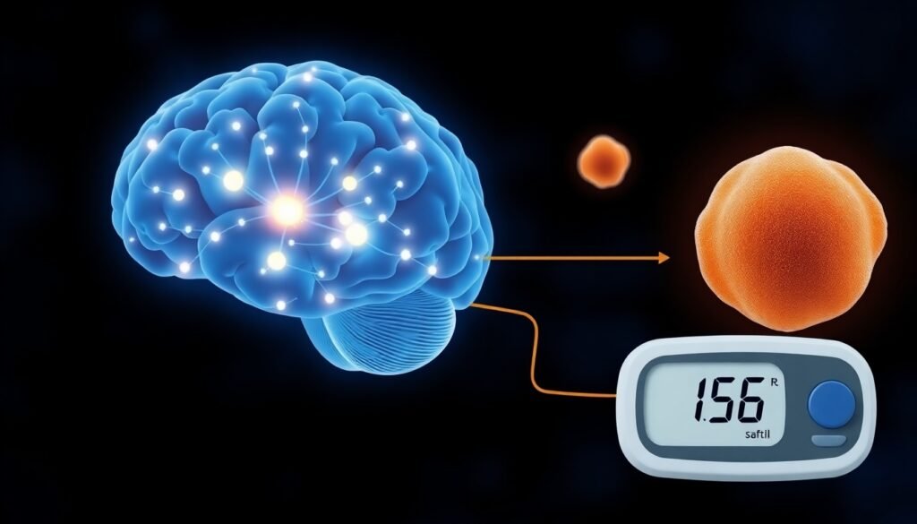
So, how do these neurons accomplish this feat? The study revealed that they act as conductors of a process called lipolysis. When activated, the VMHCckbr neurons send signals that direct the body to break down fat. This process releases glycerol, a component of fat molecules, into the bloodstream. The glycerol then travels to the liver, where it is converted into the glucose the body needs to stay powered through the night. To further confirm this mechanism, the researchers artificially activated the VMHCckbr neurons in the mice and observed a corresponding increase in glycerol levels, validating the link between these brain cells and fat metabolism.
A Potential Link to Prediabetes
The implications of this discovery extend beyond basic biology and into the realm of metabolic disease. One of the puzzling characteristics of prediabetes is an increase in lipolysis, or fat breakdown, during the night. This leads to an overproduction of glucose, contributing to the higher-than-normal blood sugar levels that define the condition.
The findings from Dr. Affinati’s team offer a compelling explanation. They suggest that in individuals with prediabetes, these VMHCckbr neurons might be overactive. This hyperactivity could be driving the excessive lipolysis, effectively flooding the body with raw materials for glucose production at a time when it isn’t needed. This constant, low-level overproduction of glucose could be a key factor in the progression from a healthy metabolic state to prediabetes and eventually type 2 diabetes.
A More Complex System
This research fundamentally shifts our understanding of how the brain manages metabolism. It moves away from a simple, binary model of an


