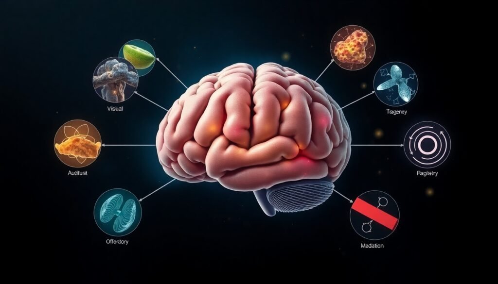
New research reveals how the locus coeruleus acts as a ‘memory reset,’ segmenting our experiences into distinct events. But what happens when chronic stress throws this system out of balance?
Life unfolds in a continuous, seamless flow of moments. Yet, when we look back, our memories aren’t a single, unbroken recording. Instead, they are a collection of distinct stories, chapters, and episodes. We remember our first day of school, a memorable vacation, or a significant conversation as individual events, not as a continuous stream of consciousness. This mental organization gives our life’s narrative shape and coherence, much like punctuation gives structure to a sentence. It helps us understand what happened, when it happened, and why it was important.
A fundamental question in neuroscience has been: how does the brain know when to end one memory and begin the next? How does it insert the commas, periods, and paragraph breaks into our personal history? One might assume such a monumental task would require vast neural real estate. However, a groundbreaking study published in the journal Neuron reveals that this critical function is orchestrated by a surprisingly small, yet incredibly powerful, region deep within the brainstem.
Researchers from UCLA and Columbia University have identified a tiny cluster of neurons called the locus coeruleus (LC) as the brain’s primary “memory reset button.” This small region, central to the brain’s arousal system, appears to be responsible for punctuating our thoughts and memories, signaling when one meaningful event has concluded and a new one should be logged.
Designing an Experiment to Find the ‘Reset’ Signal
To uncover the mechanics of this process, the scientific team, led by UCLA psychology professor David Clewett, devised a clever experiment. They recruited 32 volunteers and had them view a series of pictures of neutral objects while inside an fMRI scanner, a machine that measures brain activity by tracking changes in blood flow.
The key was to manipulate the context in which the participants saw these objects. The researchers created distinct “events” using simple auditory cues. For a period, a series of pure tones was played into the participant’s right ear, creating a stable, coherent context. Then, the tone would suddenly switch to the left ear and change in pitch. This shift acted as an “event boundary,” signaling a change in the environment.
Throughout this process, the researchers were monitoring two key things. First, the fMRI scanner was focused on activity in the locus coeruleus and the hippocampus, a brain region famous for its central role in forming new memories and tracking contextual details like time and place. Second, they simultaneously measured the dilation of the participants’ pupils. It’s known that our pupils dilate slightly in response to new or significant events, a response that is tightly linked to bursts of activity in the locus coeruleus. This pupillometry data provided a valuable cross-check, confirming that the fMRI signals were indeed capturing the activity of this tiny brainstem nucleus.
Connecting the Dots: From Brain Activity to Memory
The results were compelling. As predicted, the locus coeruleus became most active precisely at the event boundaries—the moments when the auditory tone switched. This burst of activity acted as a powerful signal that had direct consequences for memory.
To test this, the researchers later asked participants to reconstruct the order of the objects they had seen. They found that people were significantly worse at remembering the correct sequence of items that appeared on either side of an event boundary. This suggests that the burst of LC activity effectively separated the memories, filing them away as two distinct episodes rather than one continuous sequence.
Furthermore, the study revealed a direct conversation between the locus coeruleus and the hippocampus. Stronger LC activation at a boundary predicted larger changes in the patterns of activity within the hippocampus. As co-author Lila Davachi from Columbia University explained, it’s as if the locus coeruleus sends a critical “start signal” to the hippocampus, essentially saying, “Pay attention! We are in a new event now.” The hippocampus then gets to work mapping the structure of this new episode, creating a fresh index for a new memory.
The Complication of Chronic Stress
The locus coeruleus, however, has a dual personality. It operates in two distinct modes. The first is a rapid, “burst-like” mode that fires in response to significant events, helping to form new memories, as seen in the experiment. The second is a slower, “background” mode that regulates our general state of alertness and stress.
This is where the story takes a critical turn. What happens when the background mode is chronically overactive? Professor Clewett uses a powerful analogy: “The locus coeruleus is like the brain’s internal alarm system. But under chronic stress, this system becomes overactive. The result is like living with a fire alarm that never stops ringing, making it difficult to notice when a real fire breaks out.”
In this state of hyperarousal, the brain’s ability to detect meaningful changes is blunted. The “burst” signal that should punctuate a new event gets lost in the noise of the constant background alarm. To test this hypothesis, the researchers used an advanced imaging technique that measures neuromelanin, a pigment that accumulates in the locus coeruleus over time with repeated activation. Higher levels of neuromelanin are thought to be an indirect marker of chronic stress and hyperactivity.
Just as they suspected, participants with higher neuromelanin levels showed weaker pupil dilation and weaker LC activation in response to the event boundaries. Their brains were less sensitive to the contextual shifts, disrupting the very cues that anchor and organize new memories. This finding suggests that chronic stress can fundamentally impair our ability to structure our experiences, potentially leading to a more disorganized and fragmented memory of our lives.
Implications for Memory and Mental Health
The identification of the locus coeruleus as the conductor of memory formation is more than just a fascinating piece of neuroscience; it opens new doors for treating debilitating memory-related disorders. In conditions like Post-Traumatic Stress Disorder (PTSD) and Alzheimer’s disease, the locus coeruleus is known to be unusually hyperactive.
This research provides a clear mechanism explaining how that hyperactivity could contribute to the symptoms of these diseases. For instance, the fragmented, intrusive memories in PTSD might stem from a dysfunctional event segmentation system. By understanding this system, we can begin to develop targeted therapies. The study suggests that interventions aimed at “quieting” an overactive locus coeruleus—whether through pharmacology, mindfulness practices like slow-paced breathing, or even simple biofeedback techniques—could be a promising avenue for protecting or restoring healthy memory function.
Ultimately, this work highlights how a tiny, ancient part of our brain plays a profound role in shaping our most human quality: our ability to remember our lives as a coherent story. It’s a powerful reminder that in the intricate machinery of the brain, sometimes the smallest players have the biggest impact on how we understand and remember our world.


