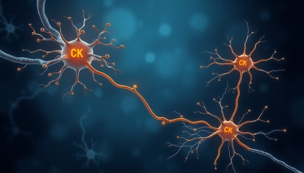
New research uncovers how a specific type of brain cell uses the neuropeptide cholecystokinin (CCK) to strengthen inhibitory signals, providing a new understanding of how the brain controls fear.
Our brains are masterpieces of plasticity, constantly remodeling themselves as we learn, remember, and experience the world. At the heart of this process are synapses, the tiny connections between neurons. For decades, neuroscientists have focused on how strengthening these connections—a process called long-term potentiation (LTP)—forms the bedrock of memory. But what about the opposite? What about the brain’s brakes? Just as important as strengthening connections is the ability to apply inhibition, to quiet down circuits and fine-tune neural communication. A new study delves into this crucial but less-understood side of brain plasticity, revealing a fascinating mechanism where specific inhibitory neurons can be powered up to weaken fear-associated memories.
This process, known as inhibitory long-term potentiation (i-LTP), is like turning up the volume on the brain’s “stop” signals. Researchers have now identified a key player in this process: a special class of inhibitory neurons that express a neuropeptide called cholecystokinin (CCK). In a series of elegant experiments, scientists have shown how these CCK-expressing neurons in the brain’s auditory cortex can not only strengthen their own inhibitory power but also empower their neighbors, ultimately leading to a measurable reduction in fear responses in mice.
Pinpointing the Master Regulator of Inhibition
The brain’s cortex contains a diverse population of inhibitory neurons, often categorized by the unique molecules they produce, such as parvalbumin (PV), somatostatin (SST), and, of course, cholecystokinin (CCK). To figure out which of these cells were responsible for i-LTP, scientists used optogenetics—a technique that uses light to control genetically modified neurons—to activate different types of inhibitory cells in the auditory cortex of mice.
First, they activated all major inhibitory neurons (GABAergic neurons) and observed a robust and lasting increase in inhibitory signals onto the principal pyramidal neurons. This confirmed that i-LTP was a real phenomenon in this brain region. The next step was to isolate the specific cell type responsible. They systematically activated each sub-class of interneuron one by one.
The results were striking. When they activated PV neurons or SST neurons alone, nothing happened. The inhibitory signals remained unchanged. But when they stimulated the CCK-expressing GABA neurons, they saw the powerful and permanent i-LTP effect once again. This discovery singled out CCK neurons as the essential drivers of this unique form of synaptic plasticity.
CCK: The Secret Ingredient for Brain Plasticity
What makes these CCK neurons so special? The researchers hypothesized that the neuropeptide CCK itself was the key ingredient. To test this, they performed another clever experiment. They went back to the PV and SST neurons—the ones that couldn’t induce i-LTP on their own—and activated them while bathing the brain tissue in an external solution containing CCK.

Suddenly, these neurons could do what was previously impossible. With the help of exogenous CCK, both PV and SST neurons were now able to trigger powerful i-LTP. This confirmed that CCK acts as a critical neuromodulator, a chemical signal that can grant other neurons the ability to strengthen their inhibitory connections. It’s not just the activity of the neuron that matters, but the chemical environment it’s in.
This finding led to an even more profound discovery about how brain circuits cooperate. The team investigated whether CCK neurons could share their special ability with their neighbors, a phenomenon known as heterosynaptic plasticity. Using a sophisticated dual-color optogenetic setup to control CCK and PV neurons independently in the same animal, they found that high-frequency stimulation of the CCK neurons not only strengthened their own synapses but also potentiated the inhibitory synapses of the nearby PV neurons. In essence, the activation of CCK neurons created a ripple effect, boosting the inhibitory tone of the entire local network. It’s a beautiful example of neural teamwork, orchestrated by a single neuropeptide.
From Brain Slices to Behavior: Weakening Fear
While these discoveries in brain slices are compelling, the ultimate test is whether this cellular mechanism has a real-world impact on behavior. To find out, the researchers turned to a classic fear-conditioning paradigm. They trained mice to associate a specific sound with a mild, unpleasant foot shock. As expected, the mice quickly learned the association and would freeze in fear upon hearing the tone alone.
This is where the CCK neurons came into play. An hour after the conditioning—a critical window for memory consolidation—the scientists used optogenetics to activate the CCK-expressing neurons in the auditory cortex of one group of mice. For comparison, they activated PV neurons in another group and had a control group with no stimulation.
The next day, they tested the mice’s memory of the fearful sound. The results were dramatic. The control mice and the mice with PV neuron activation froze for a significant amount of time, showing they still had a strong fear memory. However, the mice whose CCK neurons had been stimulated showed a significantly reduced freezing response. The activation of these specific inhibitory neurons had effectively dampened the emotional power of the traumatic memory.
A New Path for Understanding Memory and Anxiety
This study elegantly connects the dots from a single molecule (CCK) to a specific cell type, a unique form of synaptic plasticity (i-LTP), and finally, to a complex behavior (fear memory). It reveals that CCK-expressing neurons act as master regulators in the auditory cortex, capable of applying a powerful brake on neural circuits to control the strength of associative memories.
The findings open up exciting new avenues for understanding how the brain maintains a healthy balance between excitation and inhibition—a balance that is often disrupted in neurological and psychiatric conditions. By demonstrating that targeted activation of an inhibitory pathway can attenuate fear memories, this research provides a potential cellular target for developing future therapies for conditions like PTSD and anxiety disorders, where the inability to suppress fearful memories is a debilitating symptom. The brain’s dimmer switch, it turns out, may hold a key to restoring peace to a mind haunted by fear.
Reference
Huang, H. F., et al. (2025). Cholecystokinin-expressing GABA neurons elicit long-term potentiation in the cortical inhibitory synapses and attenuate sound-shock associative memory. Scientific Reports. https://doi.org/10.1038/s41598-025-17065-3


