A new study reveals that Cognitive Behavioral Therapy doesn’t just change minds—it measurably alters the structure of key brain regions involved in emotion.
For decades, the idea that psychotherapy can induce lasting change has been a cornerstone of mental health treatment. The Nobel laureate neuroscientist Eric Kandel famously proposed that for any therapy to be effective long-term, it must, on some level, physically alter the brain. While we have seen clear evidence of this with medications or treatments like electroconvulsive therapy (ECT), the structural impact of “talk therapy” has remained more elusive. Now, a groundbreaking study provides compelling evidence that Cognitive Behavioral Therapy (CBT), one of the most common forms of psychotherapy for depression, leaves a tangible mark on the brain’s architecture.
Major Depressive Disorder (MDD) is a pervasive global health issue, affecting hundreds of millions of people and standing as a leading cause of disability. Previous research has consistently linked MDD to structural changes in the brain, particularly volumetric decreases in the cortico-limbic system—a network of regions including the hippocampus and amygdala that serves as the brain’s emotional command center. While we know CBT is an effective treatment for the symptoms of depression, the question remained: does it reverse or alter these underlying physical markers? A recent study published in Translational Psychiatry set out to answer this by looking directly at the brains of patients undergoing a standard course of CBT.
A Window into the Brain During Therapy
To investigate this, researchers designed a naturalistic longitudinal study, meaning they observed patients in a real-world clinical setting over time. The team recruited 30 outpatients diagnosed with MDD who were about to begin CBT and a control group of 30 healthy individuals. Both groups underwent high-resolution structural MRI scans at two points: once before the therapy began and again after the patients had completed approximately 20 CBT sessions.
In addition to the brain scans, which measured gray matter volume (GMV), participants completed a series of clinical assessments. These tests measured the overall severity of their depressive symptoms and also evaluated a specific psychological trait called alexithymia—a difficulty in identifying and describing one’s own emotions. The researchers hypothesized that not only would CBT lead to volumetric increases in the limbic system, but that these physical changes would be more closely tied to improvements in emotional awareness (alexithymia) than to a general reduction in depression scores.
What the Scans Revealed: Growth in the Brain’s Emotion Centers
First and foremost, the therapy proved effective on a clinical level. Patients reported a significant decrease in their depressive symptoms after the 20 sessions. They also showed improvement in their ability to identify their feelings, a key component of alexithymia.
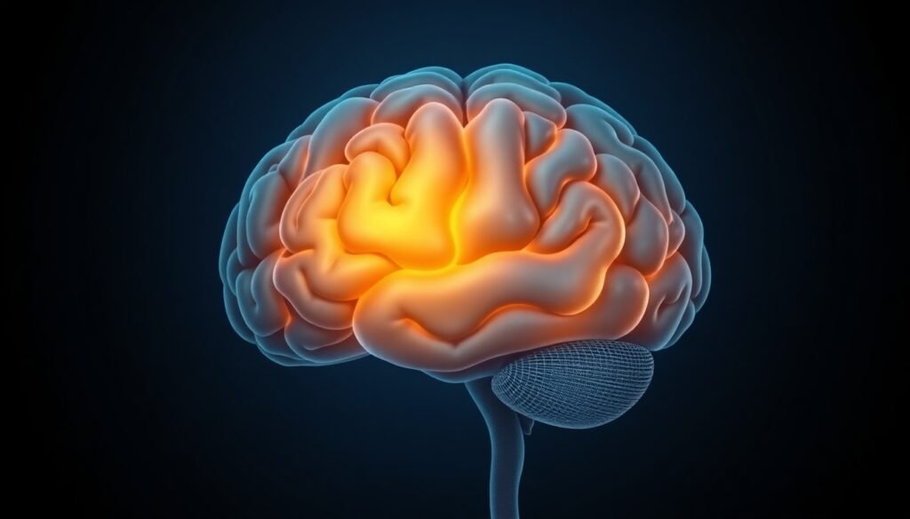
The most striking results, however, came from the MRI scans. As hypothesized, the brains of patients who underwent CBT showed significant structural changes. Specifically, the researchers observed an increase in gray matter volume in several key limbic areas:
- The Bilateral Amygdala: Both the left and right amygdala—almond-shaped structures deep in the brain known for processing emotions like fear, anxiety, and pleasure—grew in volume.
- The Right Anterior Hippocampus: The front portion of the hippocampus in the right hemisphere also increased in size. This area is critically involved in emotion regulation and memory.
These findings are remarkable because they demonstrate that a non-somatic, behavior-based intervention can induce neuroplasticity in the very same regions affected by depression and targeted by other biological treatments. The growth in the amygdala and hippocampus suggests that CBT may help strengthen the brain’s capacity for emotional processing and regulation.

Interestingly, the study also uncovered an unexpected result: a decrease in gray matter volume in the right posterior hippocampus. This part of the hippocampus is more associated with cognitive functions like spatial memory. The researchers note that this finding is puzzling and requires further investigation, as it could be related to cognitive aspects of depression that weren’t measured in the study.
The Missing Link: It’s Not Just About Feeling Better, It’s About Understanding Feelings
Perhaps the most nuanced and important finding of the study emerged when the scientists tried to connect the brain changes to the patients’ symptom improvements. They found no significant correlation between the growth in gray matter and the overall reduction in depressive symptoms. In other words, a bigger amygdala didn’t necessarily mean a patient felt less depressed overall.
However, they did find a direct link to alexithymia. The increase in volume in the right amygdala was positively correlated with a patient’s improved ability to identify their feelings. This suggests a specific mechanism: CBT may work by enhancing a person’s emotional self-awareness and processing skills. This cognitive change is then reflected in the physical structure of the brain regions responsible for that function. It’s a powerful demonstration that learning to better recognize and label emotions—a core component of many CBT exercises—has a direct, measurable biological correlate.
This finding underscores the complex and heterogeneous nature of depression. It isn’t a single entity with a single neurobiological cause. Instead, it’s a collection of diverse symptoms that may be tied to distinct dysfunctions in different brain circuits. The study suggests that successful therapy might not just shrink a single “depression area” in the brain, but rather bolster specific psychological functions, which in turn leads to structural remodeling in targeted regions.
Why This Research Matters
This study provides powerful, tangible evidence that psychotherapy is a biological treatment. For patients, this can be incredibly empowering, reframing therapy not as just talking about problems, but as an active process of retraining and reshaping the brain. It validates the hard work that goes into therapy and places it on equal footing with other medical interventions.
While the study has limitations—including a relatively small sample size and the lack of a depressed control group that did not receive therapy—its findings pave the way for a more sophisticated understanding of mental health treatment. Future research can build on this work to explore the long-term sustainability of these brain changes and even link specific therapeutic techniques to their neuroplastic effects.
Ultimately, this research moves us closer to a future of personalized medicine in mental health, where treatments can be tailored to target the specific neurobiological and psychological dysfunctions underlying an individual’s depression, optimizing their path to recovery.
Reference
Zwiky, E., Borgers, T., Klug, M., König, P., Schöniger, K., Selle, J., Küttner, A., Brunner, L., Leehr, E. J., Dannlowski, U., Enneking, V., & Redlich, R. (2025). Limbic gray matter increases in response to cognitive-behavioral therapy in major depressive disorder. Translational Psychiatry, 15(1), 301. https://doi.org/10.1038/s41398-025-03545-7


