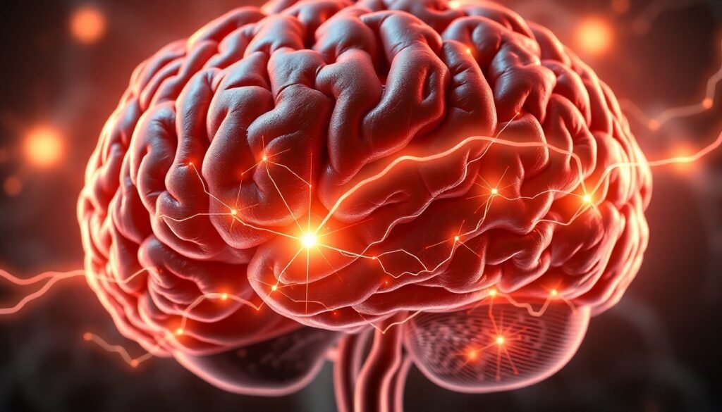
A new study reveals the popular antidepressant doesn’t just boost mood chemicals—it fundamentally changes how brain cells manage energy and rebuild connections, creating a state of enhanced flexibility.
For decades, the story of antidepressants like fluoxetine, famously known as Prozac, has been a relatively simple one: they work by increasing the levels of serotonin, the brain’s "feel-good" chemical. While this is true, it has never fully explained why these medications often take weeks to show an effect or why they don’t work for everyone. Now, groundbreaking research from a collaboration between the University of Eastern Finland and the University of Helsinki is pulling back the curtain on a much more intricate and dynamic process. It seems Prozac does more than just adjust chemical levels; it actively helps the brain rewire itself.
The new study, published in the journal Neuropsychopharmacology, suggests that fluoxetine’s true power may lie in its ability to induce neuroplasticity—the brain’s capacity to reorganize its structure, functions, or connections. The findings point to a fascinating mechanism where the drug essentially coaxes the brain out of a state of rigidness often associated with depression, creating a window of opportunity for recovery and change.
To understand this breakthrough, we first need to appreciate the brain’s architecture. Depression is increasingly seen not just as a chemical imbalance, but as a condition linked to overly rigid and inflexible neural circuits, particularly in the prefrontal cortex (PFC). The PFC is the brain’s executive suite, responsible for planning, decision-making, and moderating social behavior. In a healthy brain, this region is a bustling hub of adaptable communication. In a depressed brain, it can become stuck in negative feedback loops.
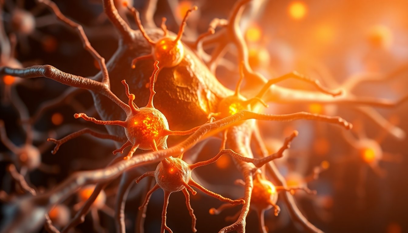
A key part of this neural landscape involves a specific type of cell: the parvalbumin-positive (PV) interneuron. Think of these cells as the brain’s traffic cops. They are a class of inhibitory neurons that help manage the flow of information, maintain network stability, and prevent brain activity from becoming chaotic. Surrounding these and other neurons is a structure called the perineuronal net (PNN). This net is a specialized part of the extracellular matrix that encases mature neurons, acting like a molecular scaffold that stabilizes synapses and, crucially, limits plasticity. While essential for maintaining stable circuits in an adult brain, this rigidity can become a barrier to recovery in conditions like depression.
The research team decided to zoom in on these specific components. Using advanced genetic tools, they investigated what happens inside the PV interneurons of the prefrontal cortex after two weeks of chronic fluoxetine treatment in an animal model. What they found was a coordinated series of changes that fundamentally altered the cells’ behavior and environment.
First, the researchers observed that the perineuronal nets around the PV cells weakened. This is a significant finding because it suggests the drug helps to literally loosen the structural constraints that keep neural circuits locked in place. By softening this scaffolding, fluoxetine may create a more permissive state for circuits to adapt and change.
But the most profound discovery was what happened inside the cells themselves. The team performed a detailed analysis of the cells’ genetic activity and found a remarkable shift in their priorities. Genes associated with energy production within the mitochondria—the tiny power plants of the cell—were significantly downregulated. This included genes for the electron transport chain, the core machinery that generates ATP, the cell’s main energy currency.
At the same time, a different set of genes was upregulated. These were genes broadly associated with plasticity: things like cytoskeletal remodeling, ion channel function, and phosphatase activity. In essence, the PV interneurons appeared to be undergoing a strategic reallocation of resources. They were spending less energy on maintaining their high-powered, stable state and investing more energy into processes that would allow them to change and form new connections.

This creates a fascinating picture of an energy-plasticity trade-off. It’s as if the antidepressant tells the brain’s traffic cops to ease up on their rigid patrols and instead focus on a city-wide roadwork project, redesigning the intersections and traffic flows. Interestingly, while the genes for energy production were turned down, the total amount of mitochondrial DNA in the cells actually increased. The researchers suggest this might be a compensatory mechanism, a way for the cell to prepare for the energy demands of future rebuilding projects, even as it dials down its current output.
So, what does this all mean for someone struggling with depression? This research helps explain why antidepressants are not a simple "happy pill." Their effect isn’t just about lifting mood directly, but about creating a state of enhanced brain flexibility. This "window of plasticity" may be why these medications are often most effective when combined with psychotherapy. The drug opens the window, and therapy provides the guidance for the brain to do the necessary rewiring—to learn new ways of thinking and break free from the rigid, negative patterns that define the depressive state.
"The findings point to a new understanding of how antidepressants may help people recover: not only by lifting mood, but by giving the brain room to rewire its circuits by altering its energy systems,” explains Senior Researcher Juzoh Umemori, the study’s leader.
Furthermore, these discoveries open up exciting new avenues for treatment. The changes in mitochondrial gene expression and the integrity of perineuronal nets could potentially serve as new biological markers to monitor whether a treatment is working. In the future, it might be possible to develop therapies that more directly target these plasticity mechanisms, perhaps leading to faster-acting or more effective treatments for depression and other mental health conditions.
Ultimately, this study moves our understanding of antidepressants beyond a simple chemical story. It paints a far more complex and hopeful picture of a dynamic process involving cellular energy, structural remodeling, and the brain’s innate potential to heal itself. Prozac, it turns out, isn’t just topping off a chemical tank; it’s helping the brain’s own mechanics get under the hood and rebuild the engine.
Reference
Umemori, J., Castrén, E., & Rantamäki, T. (2024). Chronic treatment with fluoxetine regulates mitochondrial features and plasticity-associated transcriptomic pathways in parvalbumin-positive interneurons of prefrontal cortex. Neuropsychopharmacology. https://doi.org/10.1038/s41386-024-01808-0