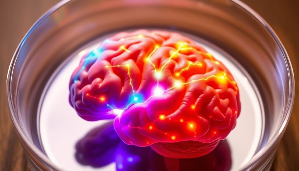
Johns Hopkins researchers have developed a multi-region brain organoid, a groundbreaking model that could revolutionize our understanding and treatment of diseases like autism and schizophrenia.
For centuries, the human brain has been the ultimate frontier of scientific exploration. Its staggering complexity, with tens of billions of neurons firing in a symphony of thought, emotion, and consciousness, makes it both a marvel and a mystery. This complexity also presents a profound challenge for researchers: how do you study the intricate workings of a living brain, especially when things go wrong? We can’t simply peek inside a person’s skull to watch a neurodevelopmental disorder unfold in real time. This fundamental limitation has long been a barrier to understanding and treating conditions that affect the entire brain, such as autism, schizophrenia, and Alzheimer’s disease.
For years, scientists have relied on animal models, but these have significant drawbacks. A mouse brain, while useful, cannot fully replicate the unique intricacies of human neurodevelopment. This gap between animal models and human reality is a major reason why drug development for neuropsychiatric disorders has a staggering failure rate, with some estimates suggesting that up to 96% of drugs fail in clinical trials. But what if we could build a better model? What if we could grow a miniature, functional human brain in a lab?
That’s precisely what a team of researchers at Johns Hopkins University has achieved. In a landmark study published in Advanced Science, they have successfully grown a novel “multi-region brain organoid” (MRBO). This isn’t just a cluster of brain cells; it’s a rudimentary, miniaturized version of a whole brain, with different regions connected and communicating with each other.
“We’ve made the next generation of brain organoids,” explained lead author Annie Kathuria, an assistant professor in JHU’s Department of Biomedical Engineering. “Most brain organoids that you see in papers are one brain region, like the cortex or the hindbrain or midbrain. We’ve grown a rudimentary whole-brain organoid.”
This breakthrough represents a monumental leap forward. To create their MRBO, Kathuria and her team took a clever, modular approach. They first grew separate organoids for different brain regions—like the cerebrum and midbrain—along with rudimentary forms of blood vessels in separate lab dishes. Then, using sticky proteins that act as a kind of biological superglue, they fused these individual components together. The result was remarkable. The distinct tissues didn’t just coexist; they began to grow together, forming connections and, crucially, producing coordinated electrical activity. They began to act as a network.

So, how closely does this lab-grown mini-brain resemble the real thing? The team found that the MRBO developed a surprisingly diverse range of neuronal cells, mirroring the complexity of a human brain at about 40 days of fetal development. In fact, about 80% of the cell types typically seen at this early stage were present and expressed in their miniature models. While vastly smaller than an adult brain—containing 6 to 7 million neurons compared to the tens of billions in a mature brain—these organoids provide an unprecedented window into the early stages of human brain development. Even more impressively, the researchers observed the formation of an early blood-brain barrier, the critical protective layer that controls what molecules can enter the brain.
The implications of this work are vast, particularly for understanding and treating complex neurological disorders. “Diseases such as schizophrenia, autism, and Alzheimer’s affect the whole brain, not just one part of the brain,” Kathuria noted. By having a model that incorporates multiple, interconnected brain regions, scientists can now study how these whole-brain disorders develop from their earliest stages.
“We need to study models with human cells if you want to understand neurodevelopmental disorders or neuropsychiatric disorders, but I can’t ask a person to let me take a peek at their brain just to study autism,” Kathuria said. “Whole-brain organoids let us watch disorders develop in real time, see if treatments work, and even tailor therapies to individual patients.”
This technology also promises to transform the landscape of drug discovery. The high failure rate of neuropsychiatric drugs is largely attributed to the poor predictive power of animal models. A drug that works in a mouse may have no effect, or even a harmful one, in a human. By providing a more accurate, human-cell-based platform for testing, these whole-brain organoids could dramatically improve the efficiency of drug screening. Researchers can apply experimental drugs directly to the organoids and observe their impact on a functional, networked system that more closely resembles a human brain.
“If you can understand what goes wrong early in development, we may be able to find new targets for drug screening,” Kathuria explained. “We can test new drugs or treatments on the organoids and determine whether they’re actually having an impact.” This could help weed out ineffective compounds early, ensuring that only the most promising candidates advance to costly and time-consuming human clinical trials.
The creation of the multi-region brain organoid is more than just a technical achievement; it’s the dawn of a new era in neuroscience. It provides a powerful tool to unravel the deepest mysteries of the brain, to understand the origins of devastating disorders, and to forge new paths toward effective treatments. While there is still a long way to go, this brain in a dish offers a tangible source of hope, bringing us one step closer to a future where the complexities of the human mind are no longer an impenetrable frontier.
Reference
Kathuria, A., et al. (2025). [Title of specific journal article not provided in source material]. Advanced Science.


