Scientists are designing custom protein fragments that intercept the toxic molecules responsible for neurodegeneration, offering a promising new therapeutic strategy.
Parkinson’s disease (PD) is a progressive neurodegenerative disorder that affects millions worldwide. At its molecular heart lies a rogue protein: alpha-synuclein (α-syn). In healthy brains, this protein exists in a soluble form, but in PD, it misfolds and clumps together into toxic, thread-like structures called fibrils. These fibrils are not just stationary plaques; they are insidious, spreading from neuron to neuron in a slow, relentless cascade that follows a predictable pattern through the brain. As they propagate, they trigger the formation of more toxic aggregates inside healthy cells and provoke neuroinflammation, leading to the devastating loss of neuronal function and the hallmark motor and cognitive symptoms of the disease.
For years, researchers have sought ways to stop this process. Now, a new study published in Nature Communications unveils an elegant and promising strategy: creating molecular decoys that intercept these toxic fibrils before they can cause harm. By designing custom protein binders, scientists have shown it’s possible to neutralize α-syn fibrils, preventing them from damaging brain cells in laboratory models.
Targeting Alpha-Synuclein’s Achilles’ Heel
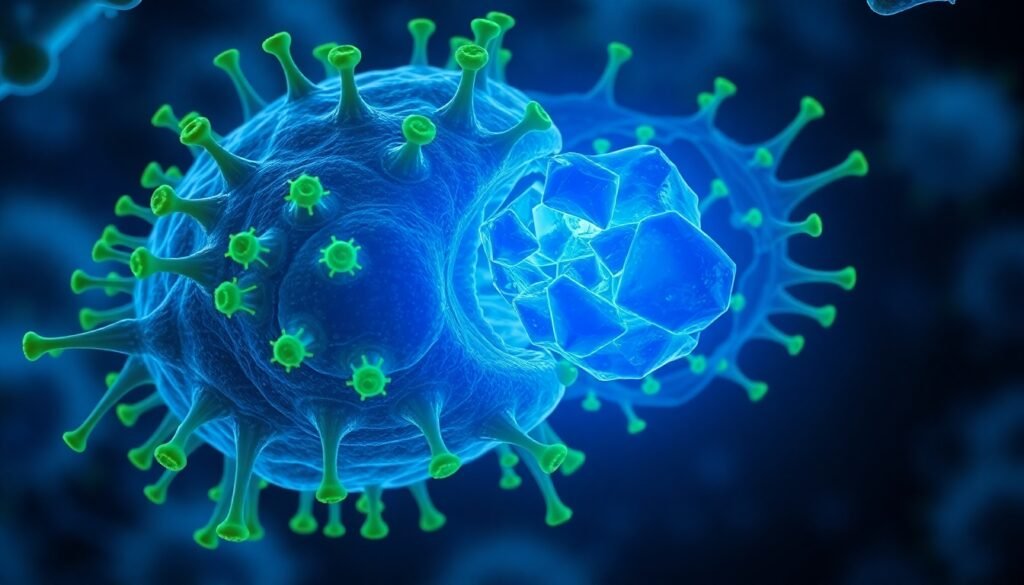
The key to the destructive power of α-syn fibrils lies in a specific segment of the protein known as the C-terminal region. This region, which is rich in negatively charged amino acids, remains exposed on the surface of the fibril. It acts like a sticky, pathological handle, allowing the fibril to latch onto various receptors on the surface of neurons and immune cells like microglia. This binding is the critical first step that enables the fibrils to invade cells, seed further aggregation, and trigger damaging inflammatory responses. The researchers behind the new study realized that if they could block this handle, they could effectively disarm the fibril.
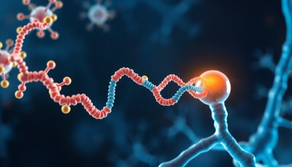
Their strategy was to fight fire with fire. They decided to use fragments of the very receptors that α-syn fibrils hijack. Specifically, they isolated the immunoglobulin-like (Ig-like) domains from two known α-syn receptors, LAG3 and RAGE. These domains, named L3D1 and vRAGE respectively, are the precise parts of the receptors that recognize and bind to the fibril’s C-terminal handle.
The hypothesis was simple: if these Ig-like domains were floating freely, could they act as decoys, binding to the α-syn fibrils and preventing them from attaching to the actual receptors on cell surfaces? The results from experiments on primary rat neurons and microglial cell lines were a resounding yes. When introduced alongside toxic α-syn fibrils, both L3D1 and vRAGE significantly reduced the amount of fibril that could adhere to the cells. This interference had profound downstream effects: it inhibited the formation of new pathological α-syn clumps inside the neurons and suppressed the release of pro-inflammatory molecules from microglia. The decoys were working.

Expanding the Arsenal and Engineering a Super-Binder
With this proof-of-concept established, the team sought to expand their arsenal of molecular decoys. Using the structure of their successful binders as a template, they identified two other Ig-like domains with high structural similarity: CD4 D1 and CAR D1. When tested, both were able to bind to α-syn fibrils, but with different affinities. CD4 D1, which had a stronger binding affinity, was also more effective at preventing fibril-induced pathology in the cell models. This finding underscored a crucial principle: the tighter the decoy’s grip on the fibril, the better its protective effect.
This led to the most compelling part of the study: rational, structure-based protein design. The team focused on the weaker binder, CAR D1, to see if they could improve it. Their analysis confirmed that the binding was primarily driven by electrostatic attraction—the positive charges on the surface of the Ig-like decoy binding to the negatively charged C-terminal handle of the α-syn fibril. The original CAR D1 had a relatively weak positive charge at its binding interface.
Using computational modeling, the researchers strategically mutated seven amino acids in CAR D1, swapping neutral or acidic residues for basic (positively charged) ones. This engineered version, dubbed CAR D1_Mut, was designed to have a much stronger electrostatic attraction to the α-syn fibril. When they produced and tested this new super-binder, the results were remarkable. CAR D1_Mut displayed a 20-fold higher binding affinity to α-syn fibrils compared to its original version, putting it on par with the highly effective CD4 D1. In cellular assays, this enhanced binding translated directly into superior protective activity, significantly inhibiting neuronal aggregation and neuroinflammation more effectively than the original CAR D1.
The Road Ahead
This research provides a powerful demonstration that engineered Ig-like binders can effectively target and neutralize the pathological activities of α-syn fibrils in cellular models. The strategy of competitive inhibition—using decoys to occupy the fibril’s binding sites—is a highly promising approach for preventing the cell-to-cell spread that drives Parkinson’s disease progression.
However, the scientists are careful to note that these findings are an early but crucial step. The experiments were conducted in vitro, using cultured cells. The next critical phase will be to validate these findings in vivo, in animal models of Parkinson’s disease. Such studies will be essential to assess the safety, stability, and therapeutic efficacy of these binders in a complex, living system. Challenges such as delivering these protein-based therapies across the blood-brain barrier and avoiding potential immune responses must also be addressed.
Despite these hurdles, this work opens a new and exciting frontier in the development of therapeutics for Parkinson’s and other related neurodegenerative diseases. By identifying a key pathological epitope and designing potent binders to target it, this study provides not only promising candidate molecules but also a blueprint for how to rationally design even better ones in the future. It is a hopeful step toward a future where the relentless progression of Parkinson’s disease can finally be brought to a halt.
Reference
Liu, C., Zhang, Y., Wang, Y., Li, Y., Zhang, Y., Li, D., … & Liu, C. (2024). Design of Ig-like binders targeting α-synuclein fibril for mitigating its pathological activities. Nature Communications, 15(1), 4755. https://doi.org/10.1038/s41467-024-49122-1


